
 Send Inquiry
Pulmonary Alveoli Model Magnified
Pulmonary Alveoli Model Magnified Specification
- Style
- Anatomic educational model
- Model No
- PA-207M
- Surface Finish
- Smooth, hand-painted
- Features
- Enlarged alveoli cluster, bronchiole branches, blood vessel detail, durable
- Power Type
- Non-electrical
- Assembly
- Ready to use, no assembly required
- Weight
- Approx. 500 grams
- Use
- Teaching and demonstration of alveolar structure
- Size
- 18 cm in height (magnified scale)
- Type
- Magnified anatomical model
- Material
- High-quality PVC plastic
- Dimensions
- Magnified view, approximate height 18 cm
- Shape
- Irregular, anatomical representation of alveoli
- Color
- Multi-colored (pink, beige, red, pale tones to indicate tissues and vessels)
- Function
- Visual aid for lung and respiratory system anatomy
- Age
- Suitable for all age groups in academic settings
- Advantage
- Highly detailed, clearly shows alveoli sac structure
- Mounting
- Mounted on Base Stand with Label
- Packing
- Supplied in Protective Carton Box
- Magnification Scale
- 130x
- Sectional View
- Provides Internal Structure and Blood Vessel Details
- Washability
- Washable and Easy to Disinfect
- Application
- Medical Training, Classroom Teaching, Demonstration
Pulmonary Alveoli Model Magnified Trade Information
- FOB Port
- Mumbai
- Payment Terms
- Cash Against Delivery (CAD), Cash on Delivery (COD), Letter of Credit (L/C), Telegraphic Transfer (T/T), Paypal, Western Union, Letter of Credit at Sight (Sight L/C), Delivery Point (DP), Days after Acceptance (DA), Cash in Advance (CID), Cheque, Cash Advance (CA)
- Supply Ability
- 50 Per Week
- Delivery Time
- 1 Week
- Sample Available
- Yes
- Sample Policy
- Sample costs shipping and taxes has to be paid by the buyer
- Packaging Details
- card board packing
- Main Export Market(s)
- Australia, North America, South America, Eastern Europe, Western Europe, Middle East, Central America, Asia, Africa
- Main Domestic Market
- All India
- Certifications
- ISO Certified product
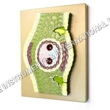
About Pulmonary Alveoli Model Magnified
- The model shows the small branches of principal bronchus:
- Section of bronchiole of no cartilage.
- The relation between pulmonary alveoli & terminal bronchiole.
- The Structure of Alveolar Sac And alveolar duct
- The capillary rete in the alveolar sapta.
Realistic Anatomy, Magnified Clarity
This pulmonary alveoli model features a highly detailed, enlarged cross-section, allowing for straightforward visualization of alveolar sacs, bronchioles, and blood vessels. Its multi-colored finish helps distinguish tissue types and vascular structures, making complex anatomical relationships more accessible for students and professionals alike.
Durable and Easy to Maintain
Made with robust, high-quality PVC plastic, this model is designed for repeated handling in academic environments. Its washable surface ensures hygiene and longevity. Simply disinfect between uses to maintain a safe and professional teaching or demonstration setting.
Seamless Teaching and Demonstration
Ideal for both classroom and medical training, this model serves as a hands-on visual aid. Its stable, labeled base and ready-to-use design mean instructors can demonstrate alveolar anatomy immediately, enhancing comprehension through direct interaction and observation.
FAQs of Pulmonary Alveoli Model Magnified:
Q: How does the magnified alveoli model assist in the understanding of lung anatomy?
A: The model magnifies the alveolar structure by 130 times, offering a clear, sectional view of internal compartments and blood vessels. This makes it easy for learners to understand the intricate relationships and functions within the respiratory system, which are otherwise difficult to observe in actual lungs.Q: What materials is the alveoli model made from, and how do I clean it?
A: The model is constructed from high-quality PVC plastic, ensuring durability and safety. Its smooth, hand-painted surface can be easily washed and disinfected with mild soap and water or suitable sanitizing wipes, supporting hygienic use in group settings.Q: When is this model typically used in academic settings?
A: This anatomical model is commonly employed during anatomy classes, medical training sessions, and demonstrations focusing on the respiratory system. It is especially useful when a detailed exploration of alveoli structure and function is required.Q: Where can the pulmonary alveoli model be used effectively?
A: The model is versatile and suitable for use in schools, colleges, universities, medical training centers, and healthcare seminars. Its portable design and protective carton box make it convenient for both stationary and mobile teaching setups.Q: What is the process for setting up and using this model during teaching?
A: No assembly is necessarythe model comes ready to use, mounted on a base with clear labeling. Instructors can immediately incorporate it into lectures, facilitating visual explanations and hands-on learning with minimal preparation time.Q: What age groups is this alveoli model designed for?
A: The model is appropriate for all age groups engaged in academic study, ranging from school students learning basic biology to university students and medical professionals seeking advanced anatomical understanding.Q: What are the primary benefits of using this magnified alveoli model?
A: Key benefits include its highly detailed structure for enhanced comprehension, durability for frequent use, ease of cleaning, and clear labeling. It effectively supports teaching and demonstration by providing a tangible, enlarged visualization of pulmonary anatomy.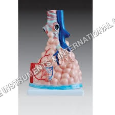

Price:
- 50
- 100
- 200
- 250
- 500
- 1000+

More Products in Anatomical Models Category
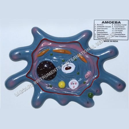
Amoeba model
Material : LLDPE (Linear LowDensity Polyethylene)
Color : MultiColor
Function : Motor skill development, Balance training
Use : Playground Equipment
Shape : Amoeba
Dimensions : 1300 mm x 440 mm x 610 mm
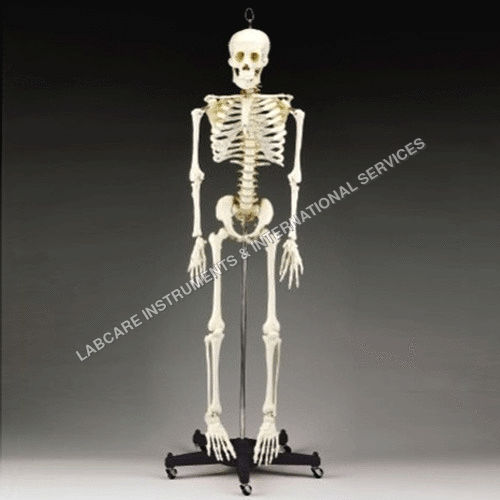
Human Skeleton model
Material : Highquality PVC plastic
Color : Natural bone color (offwhite)
Function : Demonstrates human skeletal structure
Use : Medical teaching, demonstration, study
Shape : Full adult human skeleton
Dimensions : Lifesize (approx. 180 cm height)
Development of frog model
Material : Highquality, durable PVC
Color : Multicolor lifelike finish
Function : Demonstrates frog developmental anatomy
Use : Biology teaching, anatomy studies
Shape : 3D anatomical frog
Dimensions : Approx. 20 cm x 15 cm x 8 cm
T.S. Monocot Leaf
Material : Glass slide with mounted monocot leaf section
Color : Transparent slide with stained specimen for enhanced contrast
Function : Botanical study, plant anatomy observation
Use : Educational, laboratory analysis
Shape : Rectangular
Dimensions : Standard slide dimensions (75mm x 25mm)
GOVT. APPROVED MANUFACTURER, SUPPLIER AND EXPORTER
Our Products
- Laboratory Microscope
- Hospital Furnitures
- Pharmacy Instruments
- COVID-19 SAFETY EQUIPMENTS
- Physical Instruments
- Laboratory Glasswere
- Anatomical Models
- Laboratory Glassware
- MIDWIFERY & CHILD HEALTH CARE SECTION
- Unique Products
- Chemistry Lab Equipment
- Scientific Instrument
- scientific laboratory instruments
- Physics equipments
- Hospital Equipments
- Educational Equipments kits
- ANALOG LAB TRANING MODULES
- Engineering Models & Equipments.
- Scientific Instruments
- Math catalogue Math items
- Building Furniture for School, Collegs & Office
- EDUCATIONAL WORKING MODELS
- AGRICULTURE EQUIPMENTS
- GEOGRAPHY EQUIPMENTS SECTION
- Disposible items
- The Survey Engineering Equipments Section
- Nursing College Equipments
- Fluid Machnical Lab Equipmens Labcare-Online
- Heat Transfer Lab Appartus Labcare-Online
- Labcare Entomological Equipments.
- Science and Security Equipments
- Defence Utility Equipment
- Office Equipment
- Sanitary Napkins and Diapers
- I.T.I Tools and Machines
- SCIENCE LAB EQUIPMENT
- LABTRONIKS SPECTROPHOTOMETERS
- Nebulizers


 Send Inquiry
 Send SMS
 Call Me Free
Developed and Managed by Infocom Network Private Limited.
