
 Send Inquiry
T.S. Monocot Root
T.S. Monocot Root Specification
- Surface Finish
- Smooth Glass Surface
- Features
- Labelled, Clear Sectioning, Permanent Preparation
- Power Type
- None (Passive)
- Size
- Standard Microscope Slide Size
- Model No
- TS-MONOCOT-ROOT
- Use
- Biological Studies, Laboratory Demonstration
- Assembly
- Ready to Use, No Assembly Required
- Weight
- 10-15 grams
- Style
- Educational Model
- Type
- Prepared Slide Specimen
- Material
- Glass, Prepared Slide
- Dimensions
- 44x23x4 mm approx.
- Shape
- Rectangular Slide
- Color
- Multi-Colored (Stained Tissue)
- Function
- Microscopic Observation of Monocot Root Structure
- Age
- For Students, Researchers
- Advantage
- Ready to use, Accurate representation of monocot root anatomy
- Cover Slip
- Mounted with cover slip for protection and clarity
- Magnification Support
- Suitable for low and high power objectives
- Component
- Cross-section of monocot root
- Usage Instructions
- Handle with care, for optical microscopes only
- Education Level
- Suitable for schools, colleges, and universities
- Packaging
- Individually packed in protective case
- Preservation Method
- Permanently preserved with mounting medium
T.S. Monocot Root Trade Information
- FOB Port
- MUMBAI
- Payment Terms
- Cash Against Delivery (CAD), Cash on Delivery (COD), Letter of Credit (L/C), Western Union, Paypal, Letter of Credit at Sight (Sight L/C), Telegraphic Transfer (T/T), Delivery Point (DP), Days after Acceptance (DA), Cash in Advance (CID), Cheque, Cash Advance (CA)
- Supply Ability
- 50 Per Week
- Delivery Time
- 1 Week
- Sample Available
- Yes
- Sample Policy
- Sample costs shipping and taxes has to be paid by the buyer
- Packaging Details
- card board packing
- Main Export Market(s)
- Western Europe, Asia, Australia, North America, South America, Eastern Europe, Middle East, Africa, Central America
- Main Domestic Market
- All India
- Certifications
- ISO Certified product
About T.S. Monocot Root
Accurate Monocot Root Anatomy
Every prepared slide features a true-to-life cross-section of a monocot root, stained to highlight internal structures vividly. This accurate representation helps students and researchers easily identify vascular arrangements, cortex, and epidermisvital for understanding plant biology. The permanent preparation ensures the architecture remains intact for repeated classroom and laboratory use.
Durable Glass Mounting & Clarity
Mounted under a smooth glass cover slip with a preservation medium, the specimen is protected from damage and dust. This careful mounting guarantees clear optical performance when viewed under any microscope, whether with low or high-power magnification. An individually packed case adds further protection during handling and storage.
Educational Utility Across Levels
Designed with education in mind, this prepared slide is suitable for teaching botany in schools, colleges, and universities. It serves as a valuable tool for teachers, students, and researchers, facilitating hands-on microscopic observation. Ready-to-use, labeled, and sectionedno assembly is required, making it easy to integrate into laboratory demonstrations or biological studies.
FAQs of T.S. Monocot Root:
Q: How is the T.S. Monocot Root slide preserved and protected?
A: The slide is permanently preserved using a mounting medium, and it is protected with a cover slip to maintain clarity and prevent contamination. Its packaging also includes a protective case for safe handling and storage.Q: What educational levels is this prepared slide suitable for?
A: This slide is appropriate for students and researchers in schools, colleges, and universities, making it adaptable for various grades and expertise levels in biology or plant science courses.Q: When should I use the T.S. Monocot Root slide?
A: It is ideal for use during lessons on plant anatomy, laboratory demonstrations, and research projects related to root structure and monocot plant physiology.Q: Where can the prepared slide be used?
A: The slide is designed for use in biological studios or laboratories, utilizing optical microscopes compatible with standard slide dimensions (44x23x4 mm).Q: What is the process of preparing a T.S. Monocot Root slide?
A: The monocot root section is carefully stained, mounted with a preservation medium on smooth glass, sealed with a cover slip, and labeled for identification. Each slide is then inspected for clarity and anatomical accuracy before being individually packed.Q: How should I use and handle the slide for best results?
A: Handle the slide with care to prevent breakage or contamination. Use it exclusively with optical microscopes, and avoid exposing it to chemicals or rough surfaces. The slide requires no further assembly, so its ready to use out of the box.Q: What are the main benefits of using this monocot root slide?
A: The slide offers immediate access to a clear, detailed root cross-section, supports learning through direct microscopic observation, and eliminates the need for time-consuming specimen preparationmaking it an invaluable tool in any life sciences curriculum.

Price:
- 50
- 100
- 200
- 250
- 500
- 1000+

More Products in Anatomical Models Category
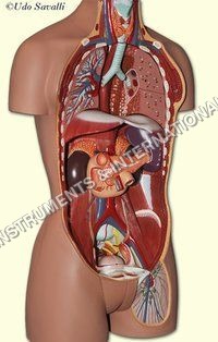
Endocrine System model
Surface Finish : Smooth, Glossy
Color : Multicolor as shown
Features : Highly Accurate, Easy to Clean, Reusable
Power Type : NonElectric
Shape : Rectangular Board with Attached 3D Glands
Size : LifeSize

Spirogyra
Surface Finish : Smooth, gelatinous surface
Color : Bright green
Features : Spiral chloroplast arrangement, rapid reproduction, nontoxic
Power Type : Requires light for growth (photosynthetic)
Shape : Filamentous, spiral chloroplasts
Size : Strands from few centimeters up to several meters
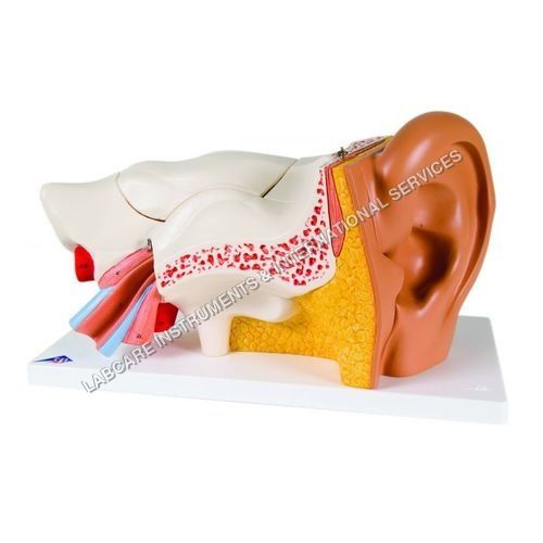
Human Ear model
Surface Finish : Smooth, hand painted, nonreflective
Color : Multicolor (realistic anatomical shades)
Features : Sectioned ear display, detachable, labeled parts
Power Type : Nonelectric
Shape : 3D lifesize human ear
Size : Lifesize
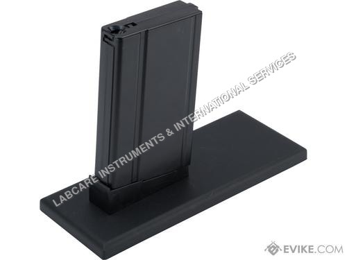
Stand for Above
Surface Finish : Smooth powder coated
Color : Black
Features : Nonslip base, adjustable clamp, high load capacity
Power Type : Nonelectric
Shape : Rectangular Base with Upright Rod
Size : Standard
GOVT. APPROVED MANUFACTURER, SUPPLIER AND EXPORTER
Our Products
- Laboratory Microscope
- Hospital Furnitures
- Pharmacy Instruments
- COVID-19 SAFETY EQUIPMENTS
- Physical Instruments
- Laboratory Glasswere
- Anatomical Models
- Laboratory Glassware
- MIDWIFERY & CHILD HEALTH CARE SECTION
- Unique Products
- Chemistry Lab Equipment
- Scientific Instrument
- scientific laboratory instruments
- Physics equipments
- Hospital Equipments
- Educational Equipments kits
- ANALOG LAB TRANING MODULES
- Engineering Models & Equipments.
- Scientific Instruments
- Math catalogue Math items
- Building Furniture for School, Collegs & Office
- EDUCATIONAL WORKING MODELS
- AGRICULTURE EQUIPMENTS
- GEOGRAPHY EQUIPMENTS SECTION
- Disposible items
- The Survey Engineering Equipments Section
- Nursing College Equipments
- Fluid Machnical Lab Equipmens Labcare-Online
- Heat Transfer Lab Appartus Labcare-Online
- Labcare Entomological Equipments.
- Science and Security Equipments
- Defence Utility Equipment
- Office Equipment
- Sanitary Napkins and Diapers
- I.T.I Tools and Machines
- SCIENCE LAB EQUIPMENT
- LABTRONIKS SPECTROPHOTOMETERS
- Nebulizers


 Send Inquiry
 Send SMS
 Call Me Free
Developed and Managed by Infocom Network Private Limited.
