
 Send Inquiry
Eye ball model
Eye ball model Specification
- Weight
- Approximately 500 grams
- Use
- Teaching aid for ophthalmology
- Model No
- EB-01
- Features
- Movable and detachable components, labeled sections
- Surface Finish
- Smooth, hand-painted
- Assembly
- Manual assembly required
- Style
- Educational anatomical model
- Size
- Life-size
- Type
- 3D Human Eye Model
- Material
- High-quality PVC plastic
- Dimensions
- Approx. 12 cm diameter
- Shape
- Spherical
- Color
- Multicolor (accurate anatomical shades)
- Function
- Demonstrates anatomical structure of human eye
- Age
- Suitable for all ages (students and professionals)
- Advantage
- Detachable parts for detailed study
- Educational Value
- Ideal for practical demonstrations, student training, and patient education
- Maintenance
- Easy to clean with mild sanitizers
- Packing
- Individual corrugated box
- Base
- Mounted on a sturdy plastic stand
- Parts Included
- 8 detachable components including cornea, iris, lens, retina
Eye ball model Trade Information
- FOB Port
- MUMBAI
- Payment Terms
- Cash Against Delivery (CAD), Cash on Delivery (COD), Cash Advance (CA), Days after Acceptance (DA), Letter of Credit (L/C), Telegraphic Transfer (T/T), Western Union, Paypal, Delivery Point (DP), Letter of Credit at Sight (Sight L/C), Cash in Advance (CID), Cheque
- Supply Ability
- 50 Per Week
- Delivery Time
- 1 Week
- Sample Available
- Yes
- Sample Policy
- Sample costs shipping and taxes has to be paid by the buyer
- Packaging Details
- card board packing
- Main Export Market(s)
- North America, South America, Eastern Europe, Middle East, Central America, Western Europe, Asia, Australia, Africa
- Main Domestic Market
- All India
- Certifications
- ISO Certified product
About Eye ball model
Comprehensive Anatomy Visualization
The EB-01 Eye Ball Model meticulously replicates the human eyes anatomical structure with life-sized accuracy. Its detachable sections, such as the cornea, iris, lens, and retina, enable in-depth demonstrations of eye anatomy, helping users visualize internal components for educational or professional sessions. The smooth, hand-painted surface provides a realistic representation essential for training and patient explanations.
Ideal for Educational and Clinical Settings
This model is specifically designed for practical demonstrations in classrooms, laboratories, clinics, and medical training centers. Medical professionals, students, and educators can all benefit from its easy-to-handle, individually packed design, making it convenient to use, transport, and set up wherever detailed eye anatomical insights are needed.
Durability and Ease of Maintenance
Constructed from high-quality PVC plastic, the Eye Ball Model ensures long-lasting use and can be easily cleaned with mild sanitizers. Its sturdy base and robust structure make it suitable for frequent handling and assembly, meeting rigorous educational and clinical demands without sacrificing clarity or precision.
FAQs of Eye ball model:
Q: How do I assemble the EB-01 Eye Ball Model?
A: The EB-01 Eye Ball Model requires manual assembly. Each of its eight detachable parts fits securely together, allowing you to easily assemble and disassemble the model for demonstration or study purposes.Q: What are the main anatomical parts included in the model?
A: The model includes eight key detachable components such as the cornea, iris, lens, and retina, all clearly labeled to promote accurate study and identification of human eye anatomy.Q: When is the Eye Ball Model most beneficial for use?
A: This model is especially valuable during practical demonstrations, classroom lessons, laboratory work, and patient education sessions where a hands-on approach aids in understanding the eyes structure.Q: Where can I use this anatomical eye model?
A: This model is suitable for use in educational institutions, medical schools, clinics, ophthalmology training centers, and even in individual study environments.Q: What process should I follow to maintain the models condition?
A: To maintain the Eye Ball Model, gently clean its surface and detachable parts with a mild sanitizer and a soft cloth. Avoid using harsh chemicals to preserve its hand-painted finish and labeled markings.Q: How does the model benefit students and professionals?
A: By providing a tactile, interactive learning experience, the Eye Ball Model helps students, trainees, and healthcare professionals understand and explain the complex structures of the human eye with greater clarity and retention.

Price:
- 50
- 100
- 200
- 250
- 500
- 1000+

More Products in Anatomical Models Category
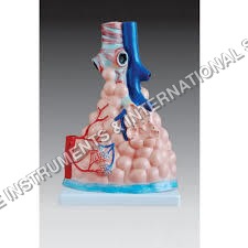
Pulmonary Alveoli Model Magnified
Shape : Irregular, anatomical representation of alveoli
Function : Visual aid for lung and respiratory system anatomy
Material : Highquality PVC plastic
Use : Teaching and demonstration of alveolar structure
Type : Other, Magnified anatomical model
Size : 18 cm in height (magnified scale)
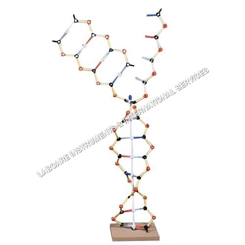
RNA model
Shape : 3D Spiral Model
Function : Demonstration of RNA Structure
Material : Highquality PVC plastic
Use : Biology Teaching, School, Laboratory, Medical Institutions
Type : Other, RNA molecular structure model
Size : Medium
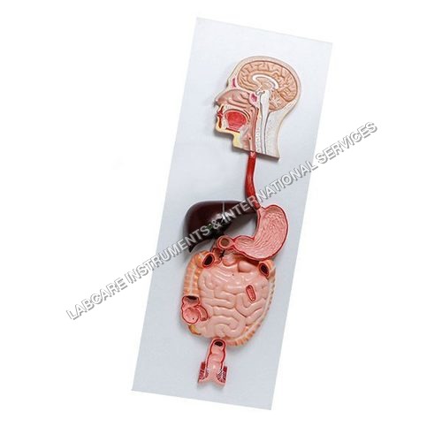
Digestive System Model
Shape : Human Digestive System Form
Function : Demonstrates Human Digestive Tract Structures
Material : HighQuality PVC Plastic
Use : Teaching Aid, Demonstration, Medical Training
Type : Other, Desktop Display Model
Size : LifeSize Approximation

Human Brain Model
Shape : Human shape
Function : bio study
Material : PVC
Use : Medical, Educational
GOVT. APPROVED MANUFACTURER, SUPPLIER AND EXPORTER
Our Products
- Laboratory Microscope
- Hospital Furnitures
- Pharmacy Instruments
- COVID-19 SAFETY EQUIPMENTS
- Physical Instruments
- Laboratory Glasswere
- Anatomical Models
- Laboratory Glassware
- MIDWIFERY & CHILD HEALTH CARE SECTION
- Unique Products
- Chemistry Lab Equipment
- Scientific Instrument
- scientific laboratory instruments
- Physics equipments
- Hospital Equipments
- Educational Equipments kits
- ANALOG LAB TRANING MODULES
- Engineering Models & Equipments.
- Scientific Instruments
- Math catalogue Math items
- Building Furniture for School, Collegs & Office
- EDUCATIONAL WORKING MODELS
- AGRICULTURE EQUIPMENTS
- GEOGRAPHY EQUIPMENTS SECTION
- Disposible items
- The Survey Engineering Equipments Section
- Nursing College Equipments
- Fluid Machnical Lab Equipmens Labcare-Online
- Heat Transfer Lab Appartus Labcare-Online
- Labcare Entomological Equipments.
- Science and Security Equipments
- Defence Utility Equipment
- Office Equipment
- Sanitary Napkins and Diapers
- I.T.I Tools and Machines
- SCIENCE LAB EQUIPMENT
- LABTRONIKS SPECTROPHOTOMETERS
- Nebulizers
- LABTRONIKS


 Send Inquiry
 Send SMS
Developed and Managed by Infocom Network Private Limited.
