
 Send Inquiry
T.S. Dicot Stem
T.S. Dicot Stem Specification
- Assembly
- Pre-assembled, Ready to Use
- Weight
- Approx. 10 g per slide
- Features
- Clearly Shows Vascular Bundles, Cortex, Epidermis, Phloem, Xylem
- Size
- 75 mm x 25 mm (Standard Slide Size)
- Use
- Biology Laboratory, Educational Purpose
- Style
- Microscope Prepared Slide
- Power Type
- Manual (Non-Electric)
- Surface Finish
- Glossy, Smooth Glass Surface
- Model No
- T.S. Dicot Stem
- Type
- Plant Anatomy Slide
- Material
- Glass Slide with Mounted Stained Plant Section
- Dimensions
- Prepared slide, standard microscope slide size (approx. 75 mm x 25 mm).
- Shape
- Rectangular Slide, Circular Section
- Color
- Stained Multicolor (Mainly Pink/Purple due to biological stains)
- Function
- Demonstration of T.S. (Transverse Section) of Dicot Stem
- Age
- Suitable for Secondary School, College, and University Use
- Advantage
- High Clarity and Accurate Representation of Dicots
- Staining Method
- Multiple stains (commonly safranin and fast green)
- Section Thickness
- 4-8 microns approx.
- Packing
- Individually packed or in box set
- Mountant Used
- Synthetic resin or Canada balsam
- Safety
- Non-toxic, Safe for classroom use
- Educational Level
- School, College, University
- Application
- Plant Anatomy, Histology Studies
- Microscope Compatibility
- Compatible with all standard light microscopes
- Image Representation
- High-definition visualization under microscope
T.S. Dicot Stem Trade Information
- FOB Port
- MUMBAI
- Payment Terms
- Cash on Delivery (COD), Delivery Point (DP), Letter of Credit (L/C), Letter of Credit at Sight (Sight L/C), Western Union, Paypal, Cash Against Delivery (CAD), Telegraphic Transfer (T/T), Days after Acceptance (DA), Cash in Advance (CID), Cheque, Cash Advance (CA)
- Supply Ability
- 50 Per Week
- Delivery Time
- 1 Week
- Sample Available
- Yes
- Sample Policy
- Sample costs shipping and taxes has to be paid by the buyer
- Packaging Details
- card board packing
- Main Export Market(s)
- Australia, North America, South America, Eastern Europe, Western Europe, Middle East, Central America, Asia, Africa
- Main Domestic Market
- All India
- Certifications
- ISO Certified product
About T.S. Dicot Stem
High-Definition Plant Anatomy Visualization
This slide offers unparalleled clarity by utilizing multiple stains that highlight the essential features of the dicot stem, such as vascular bundles and cortex regions. Students and educators can observe detailed cell structures, making it a valuable resource for engaging and accurate histology studies at every educational level.
Safe and Ready for Classroom Use
These slides are non-toxic and pre-assembled, ensuring safety for students and instructors during demonstrations. Each slide can be purchased individually or as part of a box set. With its standard dimensions and robust glass material, it fits seamlessly into any biology laboratory setup.
Compatible with All Light Microscopes
No special equipment is required to use this prepared slide. Its universal size and glossy finish guarantee easy handling and compatibility with standard light microscopes commonly found in schools, colleges, and research labs. This provides flexibility for diverse learning environments and detailed practical examination.
FAQs of T.S. Dicot Stem:
Q: How is the T.S. Dicot Stem prepared for microscopic examination?
A: The dicot stem section is cut to a thickness of approximately 48 microns, stained with biological dyes such as safranin and fast green, and mounted on a smooth glass slide using synthetic resin or Canada balsam for clear visualization.Q: What anatomical features can be observed on this slide?
A: This prepared slide clearly displays vascular bundles, cortex, epidermis, phloem, and xylem, making it suitable for detailed plant anatomy and histology studies.Q: When is this slide typically used in educational settings?
A: These slides are commonly used during biology lessons or laboratory sessions in secondary schools, colleges, and universities, particularly when studying plant structure and function.Q: Where can these prepared slides be utilized?
A: They are ideal for use in biology labs, classrooms, and research facilities. Their compatibility with standard light microscopes allows for widespread application across educational and professional environments.Q: What process is used to mount the dicot stem section on the glass slide?
A: The thin plant section is placed on the slide and fixed with a mounting medium, such as synthetic resin or Canada balsam, which preserves the specimen and provides a glossy, smooth surface for microscopic analysis.Q: How do these slides benefit biology education?
A: They provide high clarity and accurate anatomical representation, aiding effective demonstration and understanding of dicot stem structure. Their safety and ease of use also enhance the learning experience for students at various educational levels.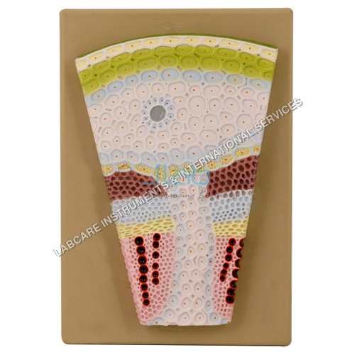

Price:
- 50
- 100
- 200
- 250
- 500
- 1000+

More Products in Anatomical Models Category
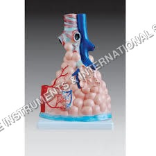
Pulmonary Alveoli Model Magnified
Advantage : Highly detailed, clearly shows alveoli sac structure
Color : Multicolored (pink, beige, red, pale tones to indicate tissues and vessels)
Features : Enlarged alveoli cluster, bronchiole branches, blood vessel detail, durable
Size : 18 cm in height (magnified scale)
Function : Visual aid for lung and respiratory system anatomy
Dimensions : Magnified view, approximate height 18 cm
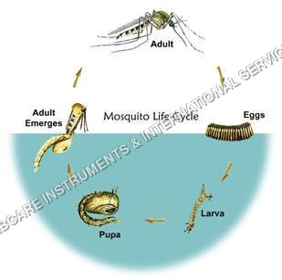
Life History of Mosquito model
Advantage : Visually Demonstrates Life Cycle Stages, Accurate Detailing
Color : Multicolor, Realistic Paint
Features : Easy to Clean, Longlasting, Clear Segmentation of Each Stage
Size : Standard Educational Size
Function : Shows detailed stages of mosquito life cycle
Dimensions : 420 mm x 300 mm
Development of frog model
Advantage : Clearly labeled internal organs, detachable parts
Color : Multicolor lifelike finish
Features : Sectional view, removable organs, detailed markings
Size : Actual frog scale
Function : Demonstrates frog developmental anatomy
Dimensions : Approx. 20 cm x 15 cm x 8 cm
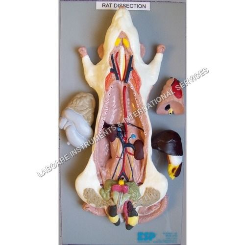
Rat Dissection model
Advantage : Reusable, hygienic alternative to real specimens
Color : Multicolor (naturalistic anatomical colors)
Features : Detachable organs, labeled internal parts, resistant to chemicals
Size : Lifesize
Function : Shows internal organs and anatomical systems of a rat
Dimensions : Approx. 30 cm length x 10 cm width x 6 cm height
GOVT. APPROVED MANUFACTURER, SUPPLIER AND EXPORTER
Our Products
- Laboratory Microscope
- Hospital Furnitures
- Pharmacy Instruments
- COVID-19 SAFETY EQUIPMENTS
- Physical Instruments
- Laboratory Glasswere
- Anatomical Models
- Laboratory Glassware
- MIDWIFERY & CHILD HEALTH CARE SECTION
- Unique Products
- Chemistry Lab Equipment
- Scientific Instrument
- scientific laboratory instruments
- Physics equipments
- Hospital Equipments
- Educational Equipments kits
- ANALOG LAB TRANING MODULES
- Engineering Models & Equipments.
- Scientific Instruments
- Math catalogue Math items
- Building Furniture for School, Collegs & Office
- EDUCATIONAL WORKING MODELS
- AGRICULTURE EQUIPMENTS
- GEOGRAPHY EQUIPMENTS SECTION
- Disposible items
- The Survey Engineering Equipments Section
- Nursing College Equipments
- Fluid Machnical Lab Equipmens Labcare-Online
- Heat Transfer Lab Appartus Labcare-Online
- Labcare Entomological Equipments.
- Science and Security Equipments
- Defence Utility Equipment
- Office Equipment
- Sanitary Napkins and Diapers
- I.T.I Tools and Machines
- SCIENCE LAB EQUIPMENT
- LABTRONIKS SPECTROPHOTOMETERS
- Nebulizers
- LABTRONIKS


 Send Inquiry
 Send SMS
Developed and Managed by Infocom Network Private Limited.
