
 Send Inquiry
T.S. Monocot stem
T.S. Monocot stem Specification
- Assembly
- No assembly required, ready to use
- Features
- Stained, labelled, sealed edges, high clarity specimen
- Surface Finish
- Smooth, polished glass with clear cover slip
- Model No
- TS-MONO-STEM-01
- Use
- Observation and study of Monocot stem T.S. under microscope
- Size
- 75 mm x 25 mm x 1 mm
- Weight
- Approx. 10-15 grams per slide
- Power Type
- Non-electrical
- Style
- Educational Microscope Slide
- Type
- Pre-mounted botanical slide
- Material
- Glass slide with preserved plant section and cover slip
- Dimensions
- Prepared slide, standard microscope slide size (75 mm x 25 mm)
- Shape
- Rectangular
- Color
- Transparent glass slide with purple/violet stained specimen
- Function
- Demonstration and teaching of Monocot stem anatomy
- Age
- Suitable for school and college level
- Advantage
- Ready-to-use, clear visualization of anatomical features, reusable
- Magnification Compatibility
- Suitable for use with standard compound microscopes (40X to 1000X)
- Preservation Method
- Long-term mounting in Canada balsam or synthetic resin
- Safety
- Non-toxic, edges sealed to prevent exposure to chemicals or breakage
- Labeling
- Printed/coded label indicating specimen name and orientation
- Staining
- Double-stained for highlighting anatomical details (commonly safranin & fast green)
- Specimen Type
- Transverse section (T.S.) of Monocot stem (typical species used: Zea mays or similar)
- Packaging
- Individually packed or in protective slide boxes
- Educational Level
- High school, undergraduate botany and biology courses
- Quality Standard
- Laboratory-grade, complies with educational supply norms
T.S. Monocot stem Trade Information
- FOB Port
- MUMBAI
- Payment Terms
- Cash Against Delivery (CAD), Cash on Delivery (COD), Telegraphic Transfer (T/T), Paypal, Letter of Credit (L/C), Western Union, Letter of Credit at Sight (Sight L/C), Delivery Point (DP), Days after Acceptance (DA), Cash in Advance (CID), Cheque, Cash Advance (CA)
- Supply Ability
- 50 Per Week
- Delivery Time
- 1 Week
- Sample Available
- Yes
- Sample Policy
- Sample costs shipping and taxes has to be paid by the buyer
- Packaging Details
- card board packing
- Main Export Market(s)
- North America, South America, Eastern Europe, Western Europe, Middle East, Asia, Central America, Australia, Africa
- Main Domestic Market
- All India
- Certifications
- ISO Certified product
About T.S. Monocot stem
Enhance Botanical Learning
This monocot stem transverse section slide offers students a clear and engaging way to study plant anatomy. The double-stained preparation accentuates vascular bundles and cellular structures, making features readily distinguishable under a microscope. Its educational design aligns with curriculum needs for high school and undergraduate courses, ensuring reliable and meaningful classroom demonstrations and individual exploration.
High-Quality and User-Friendly Design
Each slide is crafted from laboratory-grade glass with polished smooth edges and non-toxic sealing. The mounted specimen is preserved with professional standards, ensuring the slide lasts for years. Clear labeling allows for easy identification, and the slide fits all standard microscope stages, making it practical for everyday teaching, demonstration, and revision activities.
FAQs of T.S. Monocot stem:
Q: How should the T.S. Monocot Stem slide be used in classroom or laboratory settings?
A: Simply place the prepared slide onto the microscope stage. Adjust the magnification between 40X and 1000X to observe anatomical details such as vascular bundles, highlighted by the double staining. The slide is ready-to-use, requiring no setup or assembly.Q: What makes this monocot stem slide beneficial for educational purposes?
A: Its clear double-stained preparation enhances the visibility of key stem structures, facilitating easier identification and study. The slide supports curriculum requirements for high school and undergraduate biology, promoting practical understanding of monocot stem anatomy.Q: When is it recommended to use this type of microscope slide?
A: This slide is ideal for lessons covering plant anatomy, botany practicals, laboratory exams, and microscopic demonstrations related to monocotyledonous plant structure. Its reusable, making it suited for repeated classroom or laboratory use.Q: Where can the T.S. Monocot Stem slide be purchased or sourced?
A: The slide is available from laboratory equipment distributors, exporters, importers, manufacturers, service providers, suppliers, and traders across India. It may also be found in educational supply catalogs or requested directly from reputable laboratory material suppliers.Q: What is the process behind preparing this microscope slide?
A: A transverse section of monocot stem (e.g., Zea mays) is carefully sliced, double-stained with safranin and fast green to differentiate cell types, mounted in Canada balsam or synthetic resin, and sealed with a cover slip. Each slide is labeled for easy identification and preserved for long-term use.Q: How does the labeling help in practical usage?
A: Each slide features a printed or coded label indicating the specimen name and orientation, aiding quick identification and proper alignment under the microscope. This streamlines lesson flow and enhances learning efficiency during observation sessions.Q: What safety measures are incorporated into this product?
A: The slide is non-toxic, with edges securely sealed to prevent chemical exposure or accidental glass breakage. Safe for handling by students and teachers, it meets quality standards for educational laboratory materials.

Price:
- 50
- 100
- 200
- 250
- 500
- 1000+

More Products in Anatomical Models Category
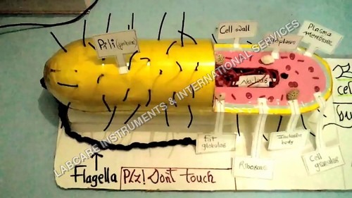
Bacteria model
Material : Highquality PVC plastic
Shape : Rodshaped bacteria
Use : Biology lab demonstration, Classroom teaching, Display
Function : Show structural details of bacteria
Color : Multicolor (Green, Yellow, Pink, Blue, etc.)
Surface Finish : Smooth, Gloss finish
Endocrine System model
Material : PVC Plastic
Shape : Rectangular Board with Attached 3D Glands
Use : Biology Lab, Medical Education, Teaching Aid
Function : Demonstrates structure and location of human endocrine glands
Color : Multicolor as shown
Surface Finish : Smooth, Glossy
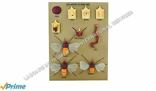
Life history of Honey bee model
Material : Durable, highquality plastic and fiber board
Shape : Rectangular display board
Use : Teaching, demonstration, and study of honey bee life cycle
Function : Helps in visualizing and understanding the honey bee life history
Color : Multicolor
Surface Finish : Glossy, easytoclean surface

Delivery process model
Material : Digital graphic (typically highresolution image or vector)
Shape : Rectangular with interconnected process boxes
Use : Visualizing and managing delivery processes
Function : Process modeling and optimization
Color : Multicolored scheme for process differentiation
Surface Finish : Glossy digital finish
GOVT. APPROVED MANUFACTURER, SUPPLIER AND EXPORTER
Our Products
- Laboratory Microscope
- Hospital Furnitures
- Pharmacy Instruments
- COVID-19 SAFETY EQUIPMENTS
- Physical Instruments
- Laboratory Glasswere
- Anatomical Models
- Laboratory Glassware
- MIDWIFERY & CHILD HEALTH CARE SECTION
- Unique Products
- Chemistry Lab Equipment
- Scientific Instrument
- scientific laboratory instruments
- Physics equipments
- Hospital Equipments
- Educational Equipments kits
- ANALOG LAB TRANING MODULES
- Engineering Models & Equipments.
- Scientific Instruments
- Math catalogue Math items
- Building Furniture for School, Collegs & Office
- EDUCATIONAL WORKING MODELS
- AGRICULTURE EQUIPMENTS
- GEOGRAPHY EQUIPMENTS SECTION
- Disposible items
- The Survey Engineering Equipments Section
- Nursing College Equipments
- Fluid Machnical Lab Equipmens Labcare-Online
- Heat Transfer Lab Appartus Labcare-Online
- Labcare Entomological Equipments.
- Science and Security Equipments
- Defence Utility Equipment
- Office Equipment
- Sanitary Napkins and Diapers
- I.T.I Tools and Machines
- SCIENCE LAB EQUIPMENT
- LABTRONIKS SPECTROPHOTOMETERS
- Nebulizers
- LABTRONIKS


 Send Inquiry
 Send SMS
Developed and Managed by Infocom Network Private Limited.
