
 Send Inquiry
Malarial parasite model
Malarial parasite model Specification
- Weight
- Approx. 0.8 kg
- Style
- Educational Display Model
- Power Type
- Non-electric, Manual display
- Use
- Teaching, Demonstration, Laboratory Study
- Surface Finish
- Smooth, Hand-painted detailing
- Model No
- MPM-101
- Size
- Enlarged for classroom demonstration
- Features
- Highly detailed, Easily washable, Non-toxic paint, Mounted on sturdy base
- Assembly
- Pre-assembled, Ready to use
- Type
- Biological Model
- Material
- High-quality PVC Plastic
- Dimensions
- 28 cm x 18 cm x 12 cm (approximate)
- Shape
- Three-dimensional life cycle stages
- Color
- Multicolor (as per actual parasite representation)
- Function
- Visual aid for understanding malarial parasite structure and lifecycle
- Age
- Suitable for all age groups, ideal for students
- Advantage
- Large size for clear visibility, Durable
- Resistant to Chemicals
- Yes
- Maintenance
- Wipe clean with damp cloth
- Packaging
- Carton box with thermocol padding
- Sections Displayed
- Sporozoite, Trophozoite, Schizont, and Gametocyte stages
- Educational Level
- School, College, University
- Base Type
- Mounted on demonstration platform
Malarial parasite model Trade Information
- FOB Port
- MUMBAI
- Payment Terms
- Cash Against Delivery (CAD), Cash on Delivery (COD), Letter of Credit (L/C), Paypal, Western Union, Letter of Credit at Sight (Sight L/C), Telegraphic Transfer (T/T), Delivery Point (DP), Days after Acceptance (DA), Cash in Advance (CID), Cheque, Cash Advance (CA)
- Supply Ability
- 50 Per Week
- Delivery Time
- 1 Week
- Sample Available
- Yes
- Sample Policy
- Sample costs shipping and taxes has to be paid by the buyer
- Packaging Details
- card board packing
- Main Export Market(s)
- Australia, North America, South America, Eastern Europe, Western Europe, Middle East, Africa, Central America, Asia
- Main Domestic Market
- All India
- Certifications
- ISO Certified product
About Malarial parasite model
Established in the year 1986 at Ambala (Haryana, India), we"LABCARE INSTRUMENTS are a Sole Proprietorship firm affianced in manufacturing highly durable array of Anatomical Models, Microbiology Models, Community Medicines Models, Forensic Models New Addition and Forensic Model. We offer these products at reasonable prices and deliver these within the promised time-frame., we have been able to provide utmost satisfaction of our clients.Comprehensive Lifecycle Representation
This model vividly displays all major stages of the malarial parasiteSporozoite, Trophozoite, Schizont, and Gametocytemaking it an essential visual aid for accurately understanding the organisms development. Each three-dimensional section is color-coded to match real-life parasite features, simplifying the study of its complex lifecycle for students at various educational levels.
Ready-to-Use and Durable Construction
Crafted from robust, high-quality PVC plastic and finished with non-toxic, hand-painted detailing, the model is mounted on a strong base for stability during demonstrations. Arriving fully assembled and packaged with thermocol padding in a secure carton box, it is resistant to chemical spills and easy to clean, ensuring both safety and durability for frequent handling in classroom or laboratory environments.
Ideal for Educational Institutions
Designed for school, college, and university use, this oversized, highly detailed malarial parasite display model benefits educators and learners alike. Its large size and clarity allow everyone in the classroom or laboratory to observe crucial structural details easily, enhancing comprehension and engagement during lessons, demonstrations, and examinations.
FAQs of Malarial parasite model:
Q: How is the malarial parasite model used in the classroom or laboratory?
A: Teachers and students use the model as a visual aid to study the different lifecycle stages of the malarial parasiteSporozoite, Trophozoite, Schizont, and Gametocyte. Its enlarged sections and three-dimensional design make it suitable for demonstrations, group discussions, and hands-on learning in both academic and laboratory settings.Q: What educational levels is this model suitable for?
A: The model is designed for a broad range of learners, making it ideal for school, college, and university-level education. Its high level of detail and large components ensure effective teaching and clear comprehension for diverse age groups.Q: When should educators consider using this model?
A: This model is best used when teaching the biology of malaria, particularly during lessons on protozoan parasites, parasitic diseases, or human health. It is also useful in laboratory classes or exam preparations to visualize and reinforce theoretical concepts.Q: Where should the model be stored after use?
A: For optimum safety and longevity, store the model in its original carton box with thermocol padding once demonstrations are complete. This protects the model from dust, accidental damage, or chemical exposure in laboratory or classroom environments.Q: What makes this malarial parasite model advantageous over smaller or less detailed versions?
A: Its enlarged size (28 x 18 x 12 cm), multicolor hand-painted detailing, and accurately represented lifecycle stages provide enhanced visibility and clarity, making it easier for students to observe and understand critical structural features, which is less feasible with compact or simply illustrated models.Q: How should the model be cleaned and maintained?
A: Maintenance is straightforward; simply wipe the surface gently with a damp cloth. The chemical-resistant, non-toxic PVC material and smooth finish allow for easy regular cleaning without damaging the models paint or details.Q: What benefits does this model offer for visual learning?
A: By presenting the malarial parasite in a tangible, enlarged, and life-like format, the model bridges the gap between textbook diagrams and real-life understanding. It enhances student engagement, aids memory retention, and supports diverse teaching styles, ultimately promoting deeper comprehension of the parasites structure and lifecycle.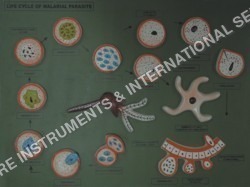

Price:
- 50
- 100
- 200
- 250
- 500
- 1000+

More Products in Anatomical Models Category
Development of frog model
Use : Biology teaching, anatomy studies
Shape : 3D anatomical frog
Material : Highquality, durable PVC
Color : Multicolor lifelike finish
Function : Demonstrates frog developmental anatomy
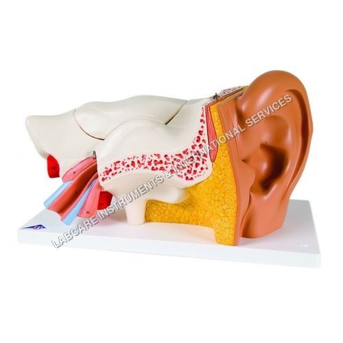
Human Ear model
Use : Medical teaching, anatomy demonstration, student learning
Shape : 3D lifesize human ear
Material : Highquality PVC plastic
Color : Multicolor (realistic anatomical shades)
Function : Demonstrates structure of external, middle, and inner ear
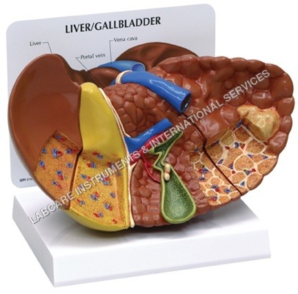
Liver model
Use : Medical Education, Demonstration, Student Learning
Shape : Threedimensional Liver Form
Material : HighQuality PVC Plastic
Color : Realistic Multicolor
Function : Displays Structure of Human Liver
Fertilization & Angiosperms
Use : Educational, teaching, botanical reference
Shape : Rectangular (if printed chart)
Material : Printed paper, chart, or digital media
Color : Multicolored (botanical illustrations)
Function : Displays process of double fertilization in flowering plants
GOVT. APPROVED MANUFACTURER, SUPPLIER AND EXPORTER
Our Products
- Laboratory Microscope
- Hospital Furnitures
- Pharmacy Instruments
- COVID-19 SAFETY EQUIPMENTS
- Physical Instruments
- Laboratory Glasswere
- Anatomical Models
- Laboratory Glassware
- MIDWIFERY & CHILD HEALTH CARE SECTION
- Unique Products
- Chemistry Lab Equipment
- Scientific Instrument
- scientific laboratory instruments
- Physics equipments
- Hospital Equipments
- Educational Equipments kits
- ANALOG LAB TRANING MODULES
- Engineering Models & Equipments.
- Scientific Instruments
- Math catalogue Math items
- Building Furniture for School, Collegs & Office
- EDUCATIONAL WORKING MODELS
- AGRICULTURE EQUIPMENTS
- GEOGRAPHY EQUIPMENTS SECTION
- Disposible items
- The Survey Engineering Equipments Section
- Nursing College Equipments
- Fluid Machnical Lab Equipmens Labcare-Online
- Heat Transfer Lab Appartus Labcare-Online
- Labcare Entomological Equipments.
- Science and Security Equipments
- Defence Utility Equipment
- Office Equipment
- Sanitary Napkins and Diapers
- I.T.I Tools and Machines
- SCIENCE LAB EQUIPMENT
- LABTRONIKS SPECTROPHOTOMETERS
- Nebulizers
- LABTRONIKS


 Send Inquiry
 Send SMS
Developed and Managed by Infocom Network Private Limited.
