
 Send Inquiry
T.S. Dicot Leaf
T.S. Dicot Leaf Specification
- Use
- Educational laboratories, teaching botany, microscopic studies
- Size
- Standard slide, 75 mm x 25 mm approx.
- Weight
- Approx. 10-15 grams per slide
- Style
- Prepared Microscope Slide
- Features
- Covers essential tissue structures like upper/lower epidermis, palisade mesophyll, spongy mesophyll, vascular bundles
- Model No
- TS-DLT-2024
- Assembly
- Mounting and coverslipping completed
- Surface Finish
- Polished glass, edge ground for safety
- Power Type
- Manual, used under light microscope
- Type
- T.S. (Transverse Section) of Dicot Leaf
- Material
- High-quality glass slide with mounted specimen
- Dimensions
- Slide size: 75 mm x 25 mm
- Shape
- Rectangular glass slide with specimen embedded
- Color
- Transparent with natural and staining contrast
- Function
- Demonstrates anatomical features of typical dicot leaf
- Age
- Suitable for all academic levels
- Advantage
- Pre-mounted, ready to use, clear observation of dicot leaf structure
- Intended Audience
- Schools, colleges, universities, research labs
- Preservation Method
- Permanently preserved with mounting medium
- Labeling
- Labeled for identification
- Quality
- Laboratory-grade; clear, undamaged specimen
- Packaging
- Individually boxed or grouped in secure case
- Specimen Origin
- Natural dicot plant leaf
- Magnification Compatibility
- 10X, 40X, 100X objective lenses
- Safety
- Edges ground for safe handling
- Cover Slip
- Included and pre-sealed
T.S. Dicot Leaf Trade Information
- FOB Port
- MUMBAI
- Payment Terms
- Cash Against Delivery (CAD), Cash on Delivery (COD), Letter of Credit (L/C), Paypal, Delivery Point (DP), Western Union, Letter of Credit at Sight (Sight L/C), Telegraphic Transfer (T/T), Days after Acceptance (DA), Cash in Advance (CID), Cheque, Cash Advance (CA)
- Supply Ability
- 50 Per Week
- Delivery Time
- 1 Week
- Sample Available
- Yes
- Sample Policy
- Sample costs shipping and taxes has to be paid by the buyer
- Packaging Details
- card board packing
- Main Export Market(s)
- Australia, Eastern Europe, Middle East, South America, Western Europe, Asia, Central America, North America, Africa
- Main Domestic Market
- All India
- Certifications
- ISO Certified product
About T.S. Dicot Leaf
Explore Dicot Leaf Anatomy Up Close
Our T.S. Dicot Leaf microscope slide allows users to observe key anatomical features such as upper and lower epidermis, palisade and spongy mesophyll layers, and vascular bundles. This slide brings textbook diagrams to life for students and researchers, delivering a crisp, undamaged specimen held in place by a secure mounting medium. The transparent and partially stained finish enhances tissue contrast for accurate microscopic analysis.
Laboratory-Grade Quality, Ready for Use
Each slide is crafted from premium glass, polished and edge-ground to ensure safe classroom and laboratory handling. The specimen is permanently sealed beneath a cover slip with precision. Packaging options include individual boxes or grouped secure cases, making storage and transport straightforward for distributors, exporters, and educational institutions.
FAQs of T.S. Dicot Leaf:
Q: How do I use the T.S. Dicot Leaf prepared slide in microscopic studies?
A: The T.S. Dicot Leaf slide can be placed on the stage of any standard light microscope and is compatible with 10X, 40X, and 100X objective lenses. Simply focus on the slide to observe the anatomical features of a dicot leaf, such as the epidermis, mesophyll tissues, and vascular bundles.Q: What benefit does the pre-mounted slide offer for educational purposes?
A: The pre-mounted and sealed slide saves setup time and prevents specimen damage, allowing students and researchers immediate, clear observation of dicot leaf anatomical structures during practical sessions.Q: When should this slide be used in teaching or research?
A: Use this prepared slide during lessons on plant structure, microscopy workshops, or botanical research sessions to demonstrate the transverse anatomy of a typical dicot leaf in fine detail.Q: Where can these prepared slides be utilized?
A: These slides are ideal for schools, colleges, universities, and research laboratoriesanywhere that botany and plant anatomy are taught or studied at academic levels.Q: What is the process of preserving and mounting the dicot leaf specimen?
A: The dicot leaf is obtained from natural sources, sectioned transversely, and permanently preserved in a mounting medium before being enclosed with a cover slip. All slides are sealed to prevent contamination and damage.Q: How is safety ensured during handling of the slide?
A: Safety is ensured by grinding and polishing the slide edges to prevent cuts, and by securely sealing the specimen under a cover slip so it does not move or fragment during use.Q: What features of the dicot leaf can be observed using this slide?
A: This slide showcases key tissue structures such as upper and lower epidermis, palisade mesophyll, spongy mesophyll, and vascular bundles, providing an in-depth view of dicot leaf anatomy.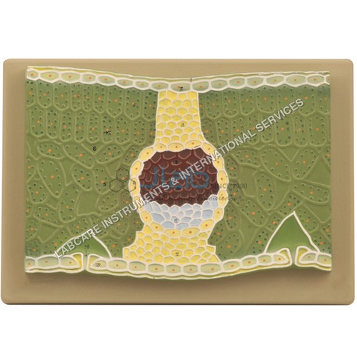

Price:
- 50
- 100
- 200
- 250
- 500
- 1000+

More Products in Anatomical Models Category

T.S. Monocot Root
Function : Microscopic Observation of Monocot Root Structure
Material : Glass, Prepared Slide
Shape : Rectangular Slide
Use : Biological Studies, Laboratory Demonstration
Advantage : Ready to use, Accurate representation of monocot root anatomy
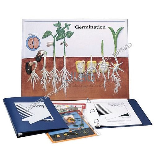
Germination & Angiosperms
Function : Study Of Germination And Angiosperms
Material : Paper
Shape : Rectangular
Use : Teaching Aid
Advantage : Clear Illustration Of Germination
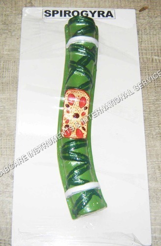
Spirogyra
Function : Produces oxygen, supports aquatic ecosystem, nutrient absorption
Material : Living algae (Spirogyra sp.)
Shape : Filamentous, spiral chloroplasts
Use : Aquatic research, water purification, education, biotechnology
Advantage : Photosynthetic, oxygengenerating, bioindicator for water health
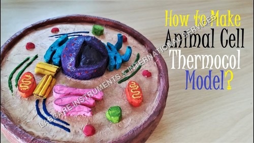
Animal cell
Function : Visual aid for biological studies
Material : PVC Plastic
Shape : Irregular (cell structure)
Use : Teaching and Demonstration
Advantage : Detailed display of animal cell organelles
GOVT. APPROVED MANUFACTURER, SUPPLIER AND EXPORTER
Our Products
- Laboratory Microscope
- Hospital Furnitures
- Pharmacy Instruments
- COVID-19 SAFETY EQUIPMENTS
- Physical Instruments
- Laboratory Glasswere
- Anatomical Models
- Laboratory Glassware
- MIDWIFERY & CHILD HEALTH CARE SECTION
- Unique Products
- Chemistry Lab Equipment
- Scientific Instrument
- scientific laboratory instruments
- Physics equipments
- Hospital Equipments
- Educational Equipments kits
- ANALOG LAB TRANING MODULES
- Engineering Models & Equipments.
- Scientific Instruments
- Math catalogue Math items
- Building Furniture for School, Collegs & Office
- EDUCATIONAL WORKING MODELS
- AGRICULTURE EQUIPMENTS
- GEOGRAPHY EQUIPMENTS SECTION
- Disposible items
- The Survey Engineering Equipments Section
- Nursing College Equipments
- Fluid Machnical Lab Equipmens Labcare-Online
- Heat Transfer Lab Appartus Labcare-Online
- Labcare Entomological Equipments.
- Science and Security Equipments
- Defence Utility Equipment
- Office Equipment
- Sanitary Napkins and Diapers
- I.T.I Tools and Machines
- SCIENCE LAB EQUIPMENT
- LABTRONIKS SPECTROPHOTOMETERS
- Nebulizers
- LABTRONIKS


 Send Inquiry
 Send SMS
Developed and Managed by Infocom Network Private Limited.
