
 Send Inquiry
T.S. Monocot Leaf
T.S. Monocot Leaf Specification
- Power Type
- Manual observation
- Use
- Educational, laboratory analysis
- Style
- Prepared microscope slide
- Surface Finish
- Smooth, polished glass
- Features
- Permanent mount, labeled, anti-fungal sealed
- Weight
- Approximately 10g
- Size
- 75mm x 25mm
- Assembly
- Ready to use, no assembly required
- Model No
- TSML-01
- Type
- Botanical microscope slide
- Material
- Glass slide with mounted monocot leaf section
- Dimensions
- Standard slide dimensions (75mm x 25mm)
- Shape
- Rectangular
- Color
- Transparent slide with stained specimen for enhanced contrast
- Function
- Botanical study, plant anatomy observation
- Age
- Suitable for all ages above 12 years
- Advantage
- Ready-to-use, clear visualization of monocot leaf structure
- Packaging
- Individual slide case for safety
- Compatibility
- Fits all standard light microscopes
- Labeling
- Printed label indicating specimen name
- Shelf Life
- Long-term, permanent preparation
- Staining
- Safranin and fast green for cellular differentiation
- Mounting Medium
- Canada balsam or equivalent
- Specimen Origin
- Natural monocot leaf sample
- Section Thickness
- Approx. 5-7 microns
T.S. Monocot Leaf Trade Information
- FOB Port
- MUMBAI
- Payment Terms
- Cash Against Delivery (CAD), Cash on Delivery (COD), Letter of Credit (L/C), Letter of Credit at Sight (Sight L/C), Telegraphic Transfer (T/T), Western Union, Paypal, Delivery Point (DP), Days after Acceptance (DA), Cash in Advance (CID), Cheque, Cash Advance (CA)
- Supply Ability
- 50 Per Week
- Delivery Time
- 1 Week
- Sample Available
- Yes
- Sample Policy
- Sample costs shipping and taxes has to be paid by the buyer
- Packaging Details
- card board packing
- Main Export Market(s)
- Australia, North America, South America, Eastern Europe, Western Europe, Middle East, Central America, Asia, Africa
- Main Domestic Market
- All India
- Certifications
- ISO Certified product
About T.S. Monocot Leaf
High-Quality Permanent Preparation
Each monocot leaf slide features a precisely thin section mounted on smooth, polished glass for maximum clarity. Canada balsam or equivalent ensures permanent adherence, protecting specimen integrity for years. Professional staining techniques using safranin and fast green offer excellent cellular contrast, making structural features easily identifiable under a microscope.
Ready-to-Use and Safe Packaging
Slides are delivered in individual cases for safety and preservation, eliminating the need for assembly. Labeled with printed specimen details, each slide is anti-fungal sealed to prevent deterioration during storage. The durable slide fits all standard light microscopes, making it suitable for classrooms, laboratories, and botanical studies.
Ideal for Botanical Education and Research
This product is tailored for educators, students (aged 12 and above), and researchers seeking detailed observation of monocot leaf anatomy. The transparent slide, enhanced by vibrant stains, facilitates learning and examination, making it an essential tool for plant science curricula and hands-on laboratory exercises.
FAQs of T.S. Monocot Leaf:
Q: How is the monocot leaf specimen processed for this microscope slide?
A: The natural monocot leaf sample is precisely sectioned to a thickness of approximately 57 microns, stained with safranin and fast green for cell differentiation, and permanently mounted on a glass slide using Canada balsam or an equivalent medium.Q: What benefits does this prepared slide offer for botanical study?
A: This slide provides a clear, ready-to-use visualization of monocot leaf structure, enabling detailed observation of cells and tissues. Its permanent preparation ensures longevity, making it ideal for repeated laboratory or educational use.Q: Where can this T.S. Monocot Leaf slide be used?
A: It is suitable for educational institutions, laboratories, and botanical research centers, offering compatibility with all standard light microscopes for effective plant anatomy observation.Q: When should this slide be used in teaching or research settings?
A: These slides can be used during botanical study sessions, laboratory practicals, biology lessons, or whenever detailed monocot leaf structure needs to be demonstrated to students or researchers above age 12.Q: What is the process behind the cellular staining on this slide?
A: Safranin and fast green stains are applied to the leaf section to differentiate cell types and structures, enabling easier identification of anatomical features under microscopic observation.Q: How does the slides packaging ensure specimen safety and quality over time?
A: Each slide is housed in an individual case and sealed with anti-fungal protection, ensuring long-term preservation and safe handling without risk of contamination or damage.Q: What makes this product compatible with most microscopes?
A: The slide is manufactured to standard dimensions (75mm x 25mm), ensuring it fits securely into all typical light microscopes used in classrooms and laboratories for manual observation.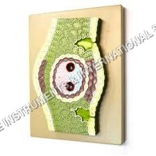

Price:
- 50
- 100
- 200
- 250
- 500
- 1000+

More Products in Anatomical Models Category

DNA model
Style : Educational Model
Use : Teaching aid, laboratory demonstration, classroom display
Advantage : Easy visualization of DNA double helix structure, colorcoded
Type : Other, 3D Molecular Model
Color : Multicolor (base pairs and backbone are color coded)
Features : Detachable parts, rotatable design, stable base
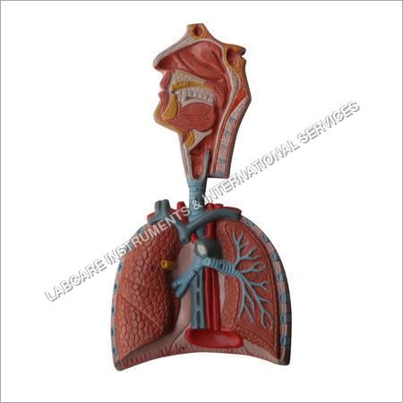
Human Respiratory System model
Style : Educational anatomical model
Use : Demonstrating human respiratory system in classrooms and laboratories
Advantage : Durable, easy to clean, accurate anatomical depiction
Type : Other, Wallmounted display model
Color : Multicolor, anatomically realistic
Features : Removable parts, numbered labels, detailed structure representation
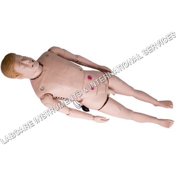
Nursing Manikin Male model
Style : Realistic human male anatomy
Use : Nursing education and training
Advantage : Durable, washable and corrosionresistant body
Type : Other, Nursing Manikin, Male Model
Color : Fleshtone
Features : Movable joints, detachable limbs, clear anatomical landmarks, easy to clean
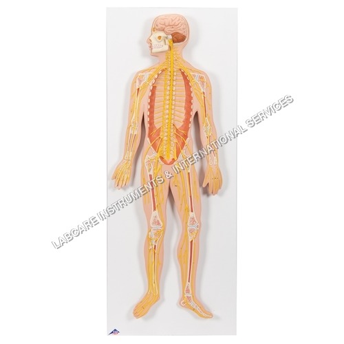
Human Nervous system model
Style : Anatomical Educational Model
Use : Medical Education, Classroom Demonstration
Advantage : Highly Detailed, Durable, Easy to Clean
Type : Other, Nervous System Model
Color : Multicolor (As Shown in Image)
Features : Demonstrates Central and Peripheral Nervous System, Detailing of Brain, Spinal Cord, and Nerves
GOVT. APPROVED MANUFACTURER, SUPPLIER AND EXPORTER
Our Products
- Laboratory Microscope
- Hospital Furnitures
- Pharmacy Instruments
- COVID-19 SAFETY EQUIPMENTS
- Physical Instruments
- Laboratory Glasswere
- Anatomical Models
- Laboratory Glassware
- MIDWIFERY & CHILD HEALTH CARE SECTION
- Unique Products
- Chemistry Lab Equipment
- Scientific Instrument
- scientific laboratory instruments
- Physics equipments
- Hospital Equipments
- Educational Equipments kits
- ANALOG LAB TRANING MODULES
- Engineering Models & Equipments.
- Scientific Instruments
- Math catalogue Math items
- Building Furniture for School, Collegs & Office
- EDUCATIONAL WORKING MODELS
- AGRICULTURE EQUIPMENTS
- GEOGRAPHY EQUIPMENTS SECTION
- Disposible items
- The Survey Engineering Equipments Section
- Nursing College Equipments
- Fluid Machnical Lab Equipmens Labcare-Online
- Heat Transfer Lab Appartus Labcare-Online
- Labcare Entomological Equipments.
- Science and Security Equipments
- Defence Utility Equipment
- Office Equipment
- Sanitary Napkins and Diapers
- I.T.I Tools and Machines
- SCIENCE LAB EQUIPMENT
- LABTRONIKS SPECTROPHOTOMETERS
- Nebulizers


 Send Inquiry
 Send SMS
Developed and Managed by Infocom Network Private Limited.
